




Quick overview
LL-37 is the only human cathelicidin, a first-line peptide of immune defence. Research focuses on biofilms, wound healing and immunomodulation.
- A natural part of human immune defence
- Studied in biofilm models where antibiotics fail
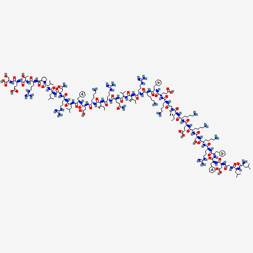
- 37 amino acids, the longest sequence in the catalogue
- Antimicrobial and regenerative research branches at once
Read more Close
Overview
Where it comes from and why it was developed
LL-37 is not a “designer” peptide in the strict sense. It is a fragment of a human immune molecule that you carry in every neutrophil, every patch of skin, and every epithelial lining. For this reason, its initial characterization did not begin in a computational design laboratory, but in blood.
In 1995, a Danish team led by Ole Sørensen (Rigshospitalet, Copenhagen) isolated from human neutrophils a molecule they named hCAP-18 (human cationic antimicrobial protein, 18 kDa). It belonged to the family of cathelicidins, an evolutionarily ancient defense system present in mammals that we share with dogs, cows, pigs, and mice. In humans, there is only one cathelicidin, encoded by the CAMP gene on chromosome 3p21.3.
Further analysis revealed that the biologically active part of hCAP-18 resides at the C-terminus, separated from the pro-domain by proteolytic cleavage. The specific enzyme responsible for this cleavage is proteinase 3 in the secretory granules of neutrophils. The resulting fragment has 37 amino acids, begins with two leucines (hence “LL”), and the term LL-37 was born.
Sørensen and colleagues published the key paper in 1997 (Blood). Since then, LL-37 has been among the most-studied human antimicrobial peptides in the literature, with more than 6,000 indexed publications.
Why the community pays so much attention to LL-37
There are three main reasons why LL-37 stands out from the crowd of peptides:
1. Antibiotic resistance. Classical antibiotics work by inhibiting specific enzymatic steps (peptidoglycan synthesis, protein synthesis, DNA replication). Bacteria have evolved resistance to them via target mutations, efflux pumps, or enzymatic degradation. LL-37 works completely differently. It attacks the physical integrity of the bacterial membrane. This mechanism is much harder to bypass by mutation, which is why LL-37 (and related peptides) represent one of the most promising directions in the development of alternatives to antibiotics for resistant strains such as MRSA, VRE, or multidrug-resistant Pseudomonas.
2. Healing of chronic wounds. In chronic venous ulcers, diabetic ulcers, and burns, the level of endogenous LL-37 in the epithelium is reduced (Heilborn 2003). Supplementation of LL-37 directly into the wound in a clinical study (Gronberg 2014) demonstrated faster closure of defects in hard-to-heal ulcers. The mechanism includes simultaneous stimulation of keratinocytes, neoangiogenesis, and antimicrobial activity, which is always needed in wound healing.
3. Immunomodulatory double-edged nature. This is the honest side that is rarely visible in marketing copy. LL-37 is not “universally beneficial.” In psoriasis, LL-37 has been found to bind self-DNA from damaged cells and form a complex that activates plasmacytoid dendritic cells via TLR9 (Lande 2007). A pro-inflammatory loop is created that sustains the chronic psoriatic state. LL-37 in psoriatic skin is therefore a mediator of disease, not a drug. You must understand this complexity when working with the peptide.
Mechanism of action, what it does at the cellular level
LL-37 is a rare case of a peptide with multiple parallel mechanisms. It is not a “one target, one effect” molecule. It is rather a modular tool of innate immunity.
Direct antimicrobial activity via membrane disruption
LL-37 is cationic (net charge +6 at physiological pH) and amphipathic (one side of the α-helix is hydrophobic, the other hydrophilic). Bacterial membranes have negatively charged phospholipids on the outer leaflet (especially phosphatidylglycerol in gram-positives, lipopolysaccharide in gram-negatives). Eukaryotic membranes have predominantly neutral phosphatidylcholine on the outer leaflet.
This is the selectivity principle. LL-37 is electrostatically attracted to the bacterial membrane, locally folds into an α-helical structure, and as an amphipathic helix inserts into the lipid bilayer. At sufficient concentration it forms transmembrane pores (the so-called “barrel-stave” or “toroidal pore” mechanism). The bacterial cell loses ionic homeostasis, lyses osmotically, and dies.
The activity spectrum is very broad:
- Gram-positive bacteria, including MRSA, VRE, Streptococcus pyogenes
- Gram-negative bacteria, including E. coli, Pseudomonas aeruginosa, Klebsiella
- Mycobacteria, including partial activity against M. tuberculosis in vitro
- Enveloped viruses: HSV-1, HSV-2, HIV, RSV, influenza, with an expected effect also against SARS-CoV-2 via envelope disruption
- Fungi, especially Candida albicans
Immunomodulation, LPS neutralization, and cytokine modulation
LL-37 does a second thing that may be even more important than direct antimicrobial activity in the context of sepsis and chronic inflammation. It binds lipopolysaccharide (LPS), the endotoxin of gram-negative bacteria, and neutralizes its ability to activate TLR4 on macrophages. This means that during systemic infection LL-37 not only kills bacteria but also dampens the pro-inflammatory response that would otherwise lead to septic shock.
Via the FPR2/ALX receptor (formyl peptide receptor 2, bound to lipoxin A4), LL-37 activates chemotaxis of:
- Neutrophils
- Monocytes
- T-lymphocytes
- Mast cells
At the same time it modulates cytokine production. In some contexts it dampens IL-6 and TNF-α (anti-inflammatory), in others (psoriasis, lupus) it increases them. Context dependence is the main feature of LL-37 that complicates therapeutic use.
Wound healing via EGFR transactivation
Tokumaru et al. (2005) showed that LL-37 activates the epidermal growth factor receptor (EGFR) in keratinocytes, but indirectly. The mechanism is interesting. LL-37 triggers release of membrane-bound HB-EGF (heparin-binding EGF-like growth factor) via activation of metalloproteinases (especially ADAM17), and the released HB-EGF then activates EGFR autocrine or paracrine. Result: migration and proliferation of keratinocytes, re-epithelialization of the wound.
Heilborn et al. (2003) completed this picture with the observation that LL-37 expression in epithelium is reduced in chronic ulcers compared to acute wounds. This led to the hypothesis that LL-37 substitution could restore healing capacity. That was confirmed in Gronberg’s Phase 1/2 study.
Stimulation of angiogenesis
Via the FPR2 receptor on endothelial cells, LL-37 stimulates endothelial cell migration and capillary tube formation. This angiogenic effect is important in healing of ischemic tissues (diabetic ulcers, burns), but in parallel raises caution in oncology models, where unwanted angiogenesis supports tumor growth.
Investigated applications
The published preclinical and clinical literature documents effects of LL-37 in the following areas:
- Antimicrobial therapy of resistant strains (MRSA, VRE, multidrug-resistant Pseudomonas)
- Healing of chronic ulcers, Phase 1/2 data (Gronberg 2014)
- Diabetic ulcers in preclinical models
- Burns, support for re-epithelialization
- Sepsis models via LPS neutralization
- Cystic fibrosis and pulmonary infections (Pseudomonas)
- IBD, Crohn’s disease, ulcerative colitis in animal models
- Atopic dermatitis, where endogenous LL-37 is reduced
- Psoriasis as a mediator of a pathological loop (in this context LL-37 is not a therapy but a target for inhibition)
- Rosacea, where elevated expression of kallikrein 5 over-produces LL-37
- Oncology research, apoptotic effects on tumor cells (context-dependent)
- Antiviral research, including hypothetical protective effect against SARS-CoV-2
Buying LL-37: what to look for
When buying LL-37, the decisive criterion is not the price but the verifiability of quality. A research peptide is only ever as good as its certificate of analysis. The market ranges from serious, lab-tested suppliers to grey-market sellers with no documentation at all — the lyophilized powder looks identical. These five criteria separate them.
1. Certificate of analysis will be supplied with the next batch
HPLC purity shows what proportion of the powder is actually LL-37. Serious suppliers document ≥ 99 % with a chromatogram. “99 % purity” without an attached chromatogram is a claim, not proof.
2. Batch-specific certificate of analysis (CoA)
The most important document. A batch-specific CoA belongs to exactly the batch you receive — with batch number, date and purity value, issued by an independent laboratory (Janoshik and similar are the industry standard). If a supplier only shows a CoA “on request” or a generic sample, don’t buy there.
3. LC-MS identity confirmation
Purity tells you how much of a substance is present; LC-MS tells you which substance it is. Via the molecular mass (4493.3 Da) it confirms this is the correct identity of LL-37, not a cheaper, mislabeled peptide.
4. Origin and EU shipping with traceability
A supplier with an EU warehouse and full batch traceability has the edge over grey imports from Asia: shorter, cooled transport and no customs risk. Molequa® ships from within the EU, typically within 3 to 5 business days — no post-Brexit customs delays.
5. Correct delivery form: lyophilizate
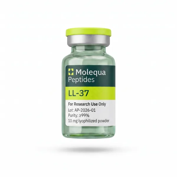
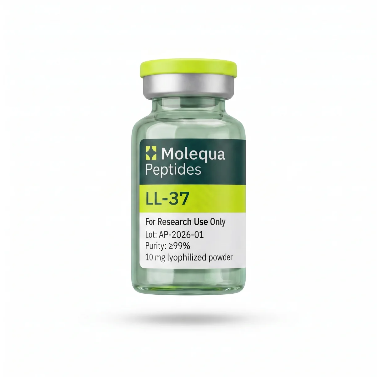
High-quality LL-37 is delivered as a lyophilizate (white powder), not as a pre-mixed solution. Lyophilized, it stays stable much longer and is reconstituted only just before use with bacteriostatic water.
Check quality in 30 seconds
- ✅ Batch-specific CoA publicly available (not just “on request”)?
- ✅ Purity proven with a chromatogram?
- ✅ LC-MS identity confirmed (mass 4493.3 Da)?
- ✅ EU warehouse and batch traceability?
- ✅ Delivered as a lyophilizate with clear storage instructions?
If all five points are met, you are buying verified material. Every Molequa® batch ships with a batch-specific certificate of analysis, and LC-MS confirmation — you can find the current CoA in the Batch test results section below.
Legal notice: LL-37 is a research peptide and not an approved medicine. It is sold exclusively for scientific laboratory research and is not intended for human or animal consumption.
Science & studies
Key publications
Sørensen O.E., et al. (1997). The human antibacterial cathelicidin, hCAP-18, is synthesized in myelocytes and metamyelocytes and localized to specific granules in neutrophils. Blood. 90(7):2796-2803. Original description of biosynthesis and localization.
Nizet V., et al. (2001). Innate antimicrobial peptide protects the skin from invasive bacterial infection. Nature. 414(6862):454-457. In vivo evidence of protective function in skin.
Heilborn J.D., et al. (2003). The cathelicidin anti-microbial peptide LL-37 is involved in re-epithelialization of human skin wounds and is lacking in chronic ulcer epithelium. J Invest Dermatol. 120(3):379-389. Key observation about chronic ulcers.
Tokumaru S., et al. (2005). Induction of keratinocyte migration via transactivation of the epidermal growth factor receptor by the antimicrobial peptide LL-37. J Immunol. 175(7):4662-4668. EGFR transactivation mechanism.
Lande R., et al. (2007). Plasmacytoid dendritic cells sense self-DNA coupled with antimicrobial peptide. Nature. 449(7162):564-569. Double-edged nature of LL-37 in psoriasis.
Carretero M., et al. (2008). In vitro and in vivo wound healing-promoting activities of human cathelicidin LL-37. J Invest Dermatol. 128(1):223-236. Comprehensive validation of the healing effect.
Gronberg A., et al. (2014). Treatment with LL-37 is safe and effective in enhancing healing of hard-to-heal venous leg ulcers: a randomized, placebo-controlled clinical trial. Wound Repair Regen. 22(5):613-621. Phase 1/2 clinical trial.
Detailed study breakdowns
Study 1: Sørensen 1997, original characterization
Citation: Sørensen O.E., Follin P., Johnsen A.H., et al. The human antibacterial cathelicidin, hCAP-18, is synthesized in myelocytes and metamyelocytes and localized to specific granules in neutrophils. Blood. 1997;90(7):2796-2803.
What they did: Isolation of hCAP-18 from human neutrophils, sequencing, identification of site of synthesis and granular localization. Methods used: chromatography, mass spectrometry, bone marrow immunohistochemistry, immunoelectron microscopy of neutrophils, in vitro cleavage by proteinase 3, and characterization of the C-terminal LL-37 fragment.
What they found:
- hCAP-18 is synthesized during the myelocyte and metamyelocyte stage of neutrophil development in bone marrow.
- It is localized to specific granules (secondary granules), not primary azurophilic granules.
- Upon degranulation, hCAP-18 is released extracellularly and proteinase 3 (from azurophilic granules) proteolytically cleaves it into the pro-domain and the active C-terminal LL-37 fragment.
- LL-37 displays antimicrobial activity against E. coli and Staphylococcus aureus at micromolar concentrations.
- Full-length hCAP-18 (without cleavage) is biologically inactive.
Why it matters: This study defined the biology of LL-37 in the human body. Without Sørensen’s discovery, we would not know that LL-37 is a “hidden” precursor in the granular system of neutrophils, activated by controlled enzymatic cleavage. All subsequent research, including therapeutic attempts to deliver synthetic LL-37 directly without dependence on proteinase 3, builds on this insight.
Study 2: Nizet 2001, in vivo protective function in skin
Citation: Nizet V., Ohtake T., Lauth X., et al. Innate antimicrobial peptide protects the skin from invasive bacterial infection. Nature. 2001;414(6862):454-457.
What they did: They used knock-out mice for cathelicidin (CRAMP, the murine equivalent of LL-37), exposed them to cutaneous inoculation with group A streptococcus (Streptococcus pyogenes), and compared them with wild-type mice. They assessed lesion size, bacterial load, and histology.
What they found:
- Knock-out mice developed markedly larger and more invasive cutaneous lesions than wild-type controls.
- Bacterial load in local tissues was 5- to 10-fold higher in knock-out mice.
- In vitro, bacteria isolated from knock-out mice had the same sensitivity to CRAMP as reference strains. The defect was therefore in host defense, not in the pathogen.
- Supplementation of exogenous CRAMP into lesions partially restored protection in knock-out mice.
Why it matters: This was the first clear in vivo evidence that cathelicidin is required for normal skin defense against bacterial infection. It elevated LL-37 (and CRAMP in mice) from the role of “interesting in vitro molecule” to that of a key component of innate immunity. The knock-out model remains the gold standard for studying the roles of AMPs in various tissues.
Study 3: Heilborn 2003, LL-37 deficit in chronic ulcers
Citation: Heilborn J.D., Nilsson M.F., Kratz G., et al. The cathelicidin anti-microbial peptide LL-37 is involved in re-epithelialization of human skin wounds and is lacking in chronic ulcer epithelium. J Invest Dermatol. 2003;120(3):379-389.
What they did: Immunohistochemical analysis of biopsies from acute wounds (surgical wounds 3 to 7 days post-incision) and chronic wounds (venous ulcers, diabetic ulcers) in human patients. In parallel, an in vitro experiment in which they blocked LL-37 in an ex vivo skin wound model and assessed the effect on re-epithelialization.
What they found:
- In acute wounds, LL-37 is strongly expressed in migrating keratinocytes at the wound edge where re-epithelialization is actively occurring.
- In chronic ulcers, LL-37 is nearly absent from the epithelium, despite increased bacterial colonization (which would normally induce its expression).
- In vitro blockade of LL-37 with antibody or antisense oligonucleotide stopped re-epithelialization in the ex vivo skin wound model.
- Supplementation of synthetic LL-37 into the model restored re-epithelialization in the blockade conditions.
Why it matters: Heilborn and colleagues linked the deficit of endogenous LL-37 to the pathophysiology of chronic wounds. This led to the hypothesis that substitution with synthetic LL-37 could be a therapeutic strategy for venous ulcers, diabetic ulcers, and other hard-to-heal defects. That hypothesis was clinically validated ten years later in Gronberg’s Phase 1/2 study.
Study 4: Tokumaru 2005, EGFR transactivation in keratinocytes
Citation: Tokumaru S., Sayama K., Shirakata Y., et al. Induction of keratinocyte migration via transactivation of the epidermal growth factor receptor by the antimicrobial peptide LL-37. J Immunol. 2005;175(7):4662-4668.
What they did: Stimulation of human keratinocytes with LL-37 in culture and assessment of:
- Migration in a scratch wound assay
- Phosphorylation of EGFR and downstream kinases (ERK1/2, Akt)
- Use of an EGFR inhibitor (AG1478) and metalloproteinase inhibitors (GM6001, TAPI-1)
- Release of HB-EGF from the cell surface
What they found:
- LL-37 at concentrations of 1 to 10 µM strongly stimulates keratinocyte migration in the scratch assay.
- LL-37 induces EGFR phosphorylation within 5 to 15 minutes, with downstream activation of ERK1/2.
- The EGFR inhibitor AG1478 completely blocks the migratory effect of LL-37.
- The ADAM metalloproteinase inhibitor (TAPI-1) also blocks migration, confirming that EGFR is activated indirectly via shedding of HB-EGF.
- LL-37 does not directly bind EGFR; the mechanism is transactivation via metalloproteinase-dependent ligand release.
Why it matters: This study revealed the concrete molecular mechanism by which LL-37 supports healing. From this it follows that LL-37 is not just an “antibiotic,” but also a growth factor modulator that engages the EGFR pathway, one of the most important pathways in epithelial regeneration. Understanding this mechanism opened the door to further studies combining LL-37 with exogenous growth factors.
Study 5: Lande 2007, double-edged nature in psoriasis
Citation: Lande R., Gregorio J., Facchinetti V., et al. Plasmacytoid dendritic cells sense self-DNA coupled with antimicrobial peptide. Nature. 2007;449(7162):564-569.
What they did: Study of psoriatic skin and an in vitro model with plasmacytoid dendritic cells (pDC). The question: why do psoriasis patients, who have elevated LL-37 levels in skin, paradoxically not have better protection from infection but instead chronic inflammation? The experiments included:
- Immunofluorescent localization of LL-37 and self-DNA in psoriatic lesions
- In vitro stimulation of pDC with combinations of LL-37 + DNA from various sources
- Measurement of IFN-α production by pDC
- TLR9 inhibition experiments
What they found:
- In psoriatic skin, LL-37 is colocalized with self-DNA released from damaged keratinocytes.
- LL-37 binds DNA via cationic interactions and transports it into pDC endosomes.
- In the endosome the DNA activates TLR9, which normally does not respond to self-DNA (because that DNA is sequestered in the nucleus and does not reach TLR9).
- Activated TLR9 triggers massive production of interferon α (IFN-α), the driving force of psoriatic inflammation.
- Without LL-37, self-DNA alone does not stimulate pDC. The combination of LL-37 + DNA is a qualitatively different signal from either alone.
Why it matters: This is a critical publication that every research user of LL-37 must know. It shows that LL-37 is not a universally “good” peptide. In some contexts (psoriasis, lupus, certain autoimmune diseases) it is a mediator of pathology, not a treatment. From the research perspective this means that context specificity is more important with LL-37 than with other peptides. You cannot simply “apply LL-37” and expect a protective effect. It depends on the tissue environment, presence of DNA, and type of immune cells.
Study 6: Carretero 2008, comprehensive validation of the healing effect
Citation: Carretero M., Escámez M.J., García M., et al. In vitro and in vivo wound healing-promoting activities of human cathelicidin LL-37. J Invest Dermatol. 2008;128(1):223-236.
What they did: A comprehensive experimental program that linked in vitro and in vivo data:
- Stimulation of human keratinocytes and fibroblasts with LL-37, measurement of proliferation, migration, and matrix protein production.
- In vivo murine model of excisional skin wounds, local application of LL-37 in a hydrogel formulation vs. control.
- Assessment of wound closure kinetics, scar tissue quality, capillary density.
- Mechanistic analysis of FPR2 receptor involvement.
What they found:
- LL-37 at concentrations of 1 to 5 µM stimulated proliferation of keratinocytes by 30 to 60 % above baseline and of fibroblasts by 20 to 40 %.
- LL-37 strongly stimulated angiogenesis in an in vitro tube formation assay with endothelial cells (HUVEC).
- In vivo: mice treated with LL-37 had faster wound closure by 25 to 35 % compared with control (days 7 and 14).
- Capillary density in healing wounds was significantly higher in the LL-37 group.
- FPR2 antagonists partially blocked the angiogenic and migratory effect of LL-37, confirming the involvement of this receptor.
Why it matters: Carretero’s study combined the cellular, mechanistic, and in vivo levels into a coherent validation of LL-37’s healing effect. It established the evidence basis for clinical translation and served as one of the main references in Gronberg’s Phase 1/2 study a few years later.
Study 7: Gronberg 2014, Phase 1/2 clinical trial in venous ulcer healing
Citation: Gronberg A., Mahlapuu M., Stahle M., Whately-Smith C., Rollman O. Treatment with LL-37 is safe and effective in enhancing healing of hard-to-heal venous leg ulcers: a randomized, placebo-controlled clinical trial. Wound Repair Regen. 2014;22(5):613-621.
What they did: Randomized, placebo-controlled, double-blind Phase 1/2 clinical trial. They enrolled patients with chronic venous leg ulcers that had not responded to standard care. Randomization: local application of synthetic LL-37 in a hydrogel formulation at three doses (0.5, 1.6, 3.2 mg/ml) vs. hydrogel placebo, twice weekly for 4 weeks. Follow-up of 8 weeks. Assessment: safety, wound size reduction, microbial colonization, local reactions.
What they found:
- No serious adverse events related to LL-37 in any group.
- Local tolerability was good; mild stinging on application was reported by <10 % of patients.
- Statistically significant acceleration of healing in the 0.5 and 1.6 mg/ml LL-37 groups vs. placebo.
- The highest dose of 3.2 mg/ml paradoxically showed a smaller effect, pointing to a bell-shaped dose-response curve (a typical phenomenon for immunomodulators).
- Bacterial load in the wound did not change significantly; the healing effect was therefore not primarily antimicrobial but regenerative.
Why it matters: This is the first and so far only published randomized clinical study of LL-37 in humans. It validated the concept of therapeutic LL-37 supplementation in the indication of chronic ulcers. The full registration process did not proceed, however. The Swedish company Pergamum AB, which sponsored the study, was later acquired by Promore Pharma, and the LL-37 clinical program underwent further restructuring. In the research context, however, Gronberg’s study remains a key safety and efficacy reference point for LL-37.
CoA, Certificate of Analysis
Quality specification (CoA with the first batch)
The values below are the release specification the first batch is tested against. The signed CoA is added as soon as it is issued.
- Purity: ≥ 98.2 % (HPLC-UV at 220 nm)
- Identity: confirmed by mass spectrometry (MS, ESI+, MW 4,493.33 Da)
- Endotoxins: < 1.0 EU/mg (LAL test, measurement of bacterial toxin contamination)
- Microbial contamination: meets USP <61>
- Residual solvents: meets ICH Q3C
- TFA residues: < 1.5 %
- Peptide content (AAA, amino acid analysis): 75 to 85 % (remainder salt and water, typical for a peptide of this size)
- Secondary structure: confirmed by CD spectroscopy (random coil in PBS, inducible α-helix in 50 % TFE)
- Related impurity profile: deletion sequences, oxidized forms < 0.5 % each
[Download CoA (PDF)], [Download SDS (PDF)]
Independent analytical laboratory (3rd-party verification). Original manufacturing CoA available upon request for B2B partners.
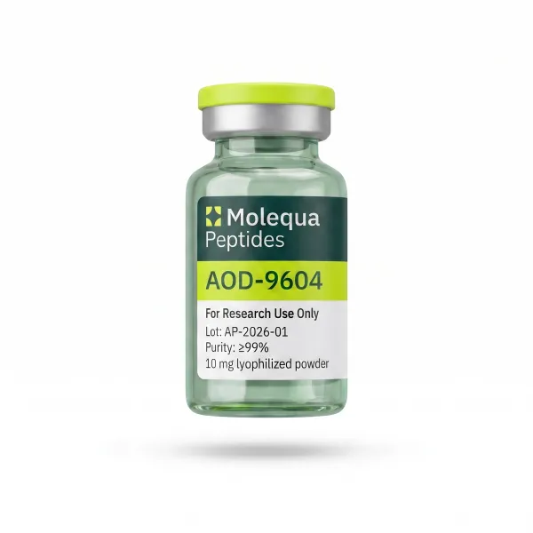
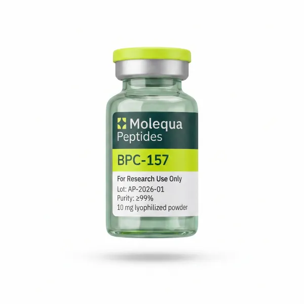
Note on synthesis: LL-37 with 37 amino acids is almost five times longer than a typical shorter peptide in our catalog (e.g. BPC-157 has 15 aa, AOD-9604 16 aa). This means significantly more synthetic steps, higher risk of deletion sequences, and more demanding purification. MOLEQUA applies a stepwise HPLC purification (first round for purity, second round for salt exchange) and verifies secondary structure for each batch by CD spectroscopy. This is why LL-37 is among our premium tier products.
Storage
Lyophilizate (dry powder before reconstitution)
- 2 years at −20 °C (freezer)
- 18 months at 2 to 8 °C (refrigerator)
- Up to 14 days at room temperature (up to 25 °C), protect from light and moisture. LL-37 is more sensitive than shorter peptides, so prolonged exposure to room temperature is not recommended.
After reconstitution (peptide in solution with bacteriostatic water)
- Up to 30 days at 2 to 8 °C, protected from light
- LL-37 in solution is more sensitive than shorter peptides. Avoid acidic conditions (pH < 4) and strongly alkaline conditions (pH > 9), which accelerate degradation. Optimal pH 5 to 7.
Practical storage rules
- Let the vial warm to room temperature (15 to 20 min) before opening. A cold vial and warm air create moisture condensation inside.
- LL-37 is an amphipathic peptide. It may to a small extent “stick” to the glass walls of the vial during prolonged storage in solution. This is a normal phenomenon; gentle swirling will release the peptide.
- Darkness is important. LL-37 does not contain tryptophan (the most sensitive aromatic amino acid), but it does contain phenylalanine and tyrosine, which also react to UV light. Store in an amber vial or box.
- Do not shake! Mechanical stress can disrupt secondary structure and promote aggregation (a typical problem for amphipathic peptides).
- The solution should remain clear. Any turbidity indicates aggregation or contamination; do not use such a sample further.
- Avoid repeated freeze-thaw cycles. If you reconstitute a larger volume, divide into aliquots and freeze once at −20 °C or −80 °C.
Combinations with peptides, frequently combined peptides
LL-37 is primarily a multifunctional peptide with both antimicrobial and regenerative effects. In research protocols it is combined with peptides that complement specific mechanisms.
BPC-157 and TB-500, wound healing and regeneration
The most common combination in the research literature on soft tissue healing. BPC-157 supports neovascularization via VEGFR2 and modulates NO synthase. TB-500 (Thymosin β4 fragment) supports cell migration and actin remodeling. LL-37 contributes an antimicrobial protective environment and EGFR-mediated keratinocyte migration. Together they cover vascularization, cell migration, and antimicrobial defense, three key components of chronic wound healing.
KPV, anti-inflammatory component for skin indications
KPV (Lys-Pro-Val, a tripeptide fragment of α-MSH) has strong anti-inflammatory effects via the melanocortin system and NF-κB inhibition. In combination with LL-37 it is being investigated for atopic dermatitis, where LL-37 supplies the antimicrobial component and KPV dampens chronic inflammation. In psoriasis the situation is more complex; KPV may be beneficial, but LL-37 may be counterproductive (Lande 2007).
Thymosin α1, immunomodulatory synergy
Thymosin α1 is a 28-amino-acid peptide with complex immunomodulatory activity, including support of T-cell maturation and activation of dendritic cells via TLR9. Combination with LL-37 is being explored for anti-infective applications (sepsis, severe infections in immunocompromised patients, certain viral infections). Both peptides modulate innate and adaptive immunity from different angles.
GHK-Cu, dermatological synergy
GHK-Cu (a tripeptide with bound copper) is well characterized in dermatology and cosmetology. It stimulates collagen and glycosaminoglycan synthesis and supports matrix remodeling. In research contexts it is combined with LL-37 for post-burn skin regeneration, anti-aging research, and support of scar healing. The mechanisms are complementary: GHK-Cu at the matrix level, LL-37 at the keratinocyte and antimicrobial protection level.
Additional note: caution in autoimmune models
In research contexts of autoimmune diseases (psoriasis, lupus, certain forms of IBD), combination of LL-37 with other immune activators is contraindicated. In these models LL-37 may contribute to pathogenesis via the TLR9-IFN-α loop (Lande 2007). Always verify the applicability of LL-37 in the specific model context first.
Key scientific figures and citations
“LL-37, the only human cathelicidin-derived antimicrobial peptide, exhibits broad-spectrum antimicrobial activity and serves as a multifunctional effector molecule of innate immunity.”
Dürr UH., Sudheendra US., Ramamoorthy A. (2006), Biochim Biophys Acta 1758(9), PubMed 16716248
Statistics from preclinical literature
- LL-37, the human cathelicidin antimicrobial peptide, 37 amino acids, sequence beginning Leu-Leu (hence the name), molecular weight 4493.3 Da
- Encoded by the CAMP gene (chromosome 3p21.3), released from the hCAP-18 protein by proteinase-3 cleavage in neutrophils
- Identified by the group of Birgitta Agerberth (Karolinska Institutet, Sweden) in 1995
- Standard experimental concentration: 1–10 μg/ml in vitro for antimicrobial assays (E. coli, S. aureus, C. albicans)
- Mechanism: direct permeabilization of the bacterial membrane (cationic amphipathic α-helix), modulation of FPR2/ALX, P2X7, GPCRs, induction of angiogenesis and re-epithelialization
- MIC against E. coli: ~5 μM (Travis et al. 2000); against S. aureus: ~10–20 μM
- 4 approved therapeutic derivatives identified in development (Pexiganan, Omiganan, Iseganan, all Phase 2/3 failed)
- Approximately 3000+ publications in PubMed (1995–2024), a cornerstone peptide of innate immunity
Reference sources (PubMed)
- Agerberth B. et al. (1995). “FALL-39, a putative human peptide antibiotic, is cysteine-free and expressed in bone marrow and testis.” Proc Natl Acad Sci USA 92(1):195–199. PubMed 7529412
- Dürr UH. et al. (2006). “LL-37, the only human member of the cathelicidin family of antimicrobial peptides.” Biochim Biophys Acta 1758(9):1408–1425. PubMed 16716248
- Vandamme D. et al. (2012). “A comprehensive summary of LL-37, the factotum human cathelicidin peptide.” Cell Immunol 280(1):22–35. PubMed 23246832
Regulatory status: LL-37 is not an approved human medicinal product in any regulatory zone (FDA, EMA, or any national medicines agency). Derivatives in development (Pexiganan/MSI-78, Omiganan) progressed through Phase 2/3 clinical trials for topical applications (diabetic foot ulcer, rosacea) but did not achieve regulatory approval. Existing data on LL-37 itself come from preclinical and in vitro literature. The product is sold strictly for laboratory scientific research (RUO).
Frequently asked questions about LL-37
These questions address the most common research-context searches about LL-37. For full technical documentation see the sections above.
What is LL-37 and what is it used for in research?
LL-37 (sequence LLGDFFRKSKEKIGKEFKRIVQRIKDFLRNLVPRTES, 37 AA, 4493 Da) is the only human cathelicidin antimicrobial peptide, derived from the hCAP-18 precursor. In research it exhibits direct bactericidal activity (via membrane permeabilisation), immunomodulation and wound-healing support. It is studied in models of chronic infections, cystic fibrosis and dermatological diseases.
What dose of LL-37 do scientists use in animal models?
In preclinical animal models LL-37 is tested topically at concentrations of 10 to 100 µg/ml or systemically at 0.5 to 5 mg/kg intraperitoneally. Clinical formulations (OP-145, ophthalmic preparations, Phase 2) test substantially lower doses to minimise cytotoxicity.
What is the difference between LL-37 and Thymosin α1?
LL-37 and Thymosin α1 are both immunomodulatory peptides, but LL-37 targets direct antimicrobial defence (innate immunity, bactericidal activity), whereas Thymosin α1 modulates adaptive immunity via T cells and TLR signalling. LL-37 has intrinsic bactericidal activity, Thymosin α1 does not.
Is LL-37 an approved medicine or research substance?
LL-37 is not an approved human medicine. Several derivatives (OP-145, omiganan) went through Phase 2/3 trials, but none achieved EMA/FDA approval. The product is sold strictly for laboratory scientific research (RUO), not for clinical use.
How is LL-37 stored?
Lyophilised LL-37 should be stored at −20 °C protected from light, stability 2 to 3 years; at 2 to 8 °C 6 months. LL-37 is amphipathic, reconstitute in sterile water without detergents, the solution is stable 14 days at 2 to 8 °C. Avoid repeated freeze/thaw cycles.
What is the half-life of LL-37 and how often is it administered in studies?
LL-37 has a short plasma half-life (~30 minutes) due to rapid proteolysis. In tissues (where it is bound to biofilm or cell membranes) the effect persists for hours. Experimental protocols use 2 to 3 daily administrations in systemic studies.
Where to buy LL-37 in the EU for scientific research?
LL-37 for scientific research in the EU is offered by Molequa® with FedEx delivery in 3 to 5 business days across the EU. The product ships lyophilised with a Certificate of Analysis (COA), HPLC purity ≥ 98.2 %. The product is strictly for laboratory scientific research (RUO).